Image Contest Exhibition
We are pleased to present the contributions to this year's Image Contest!
Apis florea colony
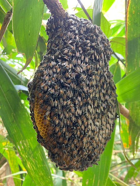
This is Apis florea, the dwarf honey bee—among the rarest and smallest Indian honey bee species we studied. Often hidden in bushes or leaf clutter and very fragile. I was captivated seeing it for the first time, with its vibrant red colors streaked with white, as if hand-painted.
- Abhinay Arra
Apis florea Comb Structure

Honey bees are remarkable builders, creating perfectly symmetrical combs using only their legs. This Apis florea comb shows clear organization: deep honey chambers at the top, smaller worker cells below, followed by larger drone cells, and queen cells at the base—a marvelous design matching bee size and function.
- Abhinay Arra
What happened to me? Can anyone help me?

CEACAM1-deficient BMDM was captured on day 0 after M-CSF treatment. The image was taken using a transmission Electron Microscope.
-Anggi Muhtar Pratama
The Birth of Platelets: A Dance of Balance

From the giant megakaryocyte, small platelets are born — fragments entwined like yin and yang, united yet independent, reminding us that life is sustained by balance.
Image was acquired with a Thunder Microscope (63x objective), scale bar 20μm. Red = nuclei, cyan = tubulin, magenta = actin.
-Gabriel Araujo
Neuron Divide: Visualizing Soma‑Axon Separation
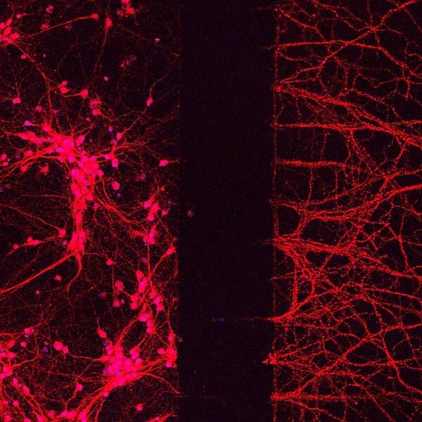
This confocal image shows mouse cortical neurons stained with DAPI (Blue), NeuN (Magenta) and Tau (Red) cultured in microfluidic chamber for 7 days. With the use of fluid pressure gradient, the axons flow through microgrooves to the axonal compartment.
-Gayatri Gandhi
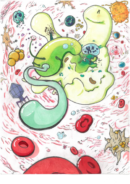
The Creation in the world of E. coli
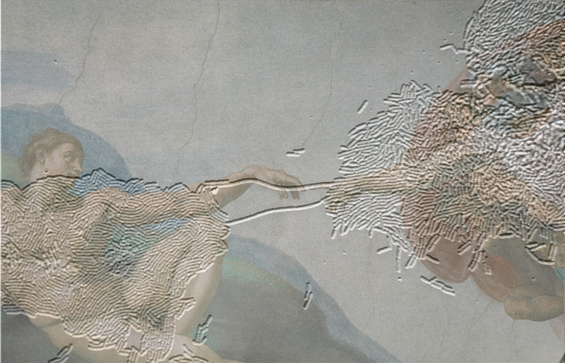
This image combines a Nikon microscope photo of genetically modified E. coli with Michelangelo’s The Creation of Adam (sourced from Wikipedia). Two large bacterial colonies are linked by elongated cells that reminded me of the famous hand gesture, which inspired me to merge them together.
-Ruilan Xu
Nanoscale Bacteriology lab, Rudolf Virchow Center for Intergrative and Translational Bioimaging, University Würzburg, 97080 Würzburg
From the Gut, with Love
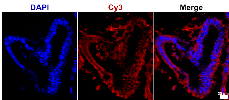
A heart-shaped mouse intestinal organoid, grown in 3D culture. Science can occasionally organize itself into art, offering as a reminder that discoveries can be both intricate and beautiful……
-Nikita Deoghare
Sphere of Promise

The process of generating a neuro-mesodermal assembloid begins with a simple sphere of human iPSCs. This cluster of cells holds immense potential, as it is capable of forming virtually any tissue or structure. Every sphere carries a promise: To enhance our understanding of human development, to uncover the mechanisms of disease, or to reveal the unexpected beauty of what can emerge from just a few cells. In the beginning, the sphere can become anything—we only have to decide what we’re ready to see in it.
- Ann-Sophie Schnell
An unhappy E. Coli strikes a dramatic pose!
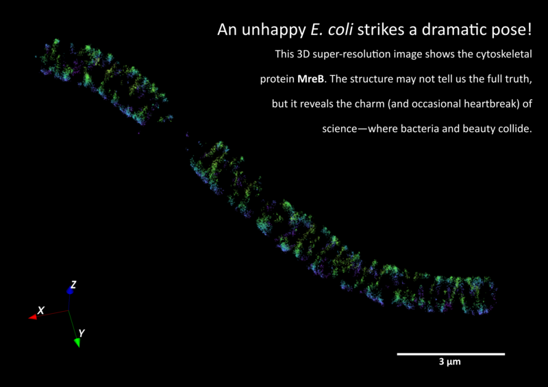
This 3D super-resolution image shows the cytoskeletal protein MreB. The structure may not tell us the full truth, but it reveals the charm (and occasional heartbreak) of science - where bacteria and beauty collide.
- Kilian Andress
When measles hits and you realize you’re the experiment
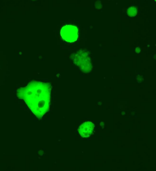
These are primary human CD4+ T cells infected with a GFP-tagged measles virus. Viral infection causes the cells to fuse with each other, resulting in this perfect “I wasn’t ready” face. Looks like even cells get surprised when viruses crash the party—a reminder that science never fails to surprise.
- Maria Grijalva Yepez