Image Contest
In the Image Contest, PhD students present fascinating moments captured from their own scientific work. The images can be taken on a variety of scientific imaging devices. In the past years the contestants allowed a broader audience to appreciate the beauty of science on any level imaginable.
Here we present the entries of this years Image Contest.
Anna Seubert - My little green cactus (Mein kleiner grüner Kacktus)
Nicole Michelle Schottmann - Schrödinger’s Myelination
Trushnal Waghmare - Ceramide moustache ghost
Denise Johnson - Inside a thrombus
Tuğçe Çimen - Cross talk between neutrophils and platelets in the inflamed microcirculation.
Alexander Veh - Help me I’m swelling!
Vibhuti Bhat - My EUREKA! moment
Ye Ouyang - Tree
Muthita Khamwong - Honeycomb architecture of Langerhans cell network in murine tail
Martina Wilhelm - Dragon of the Mind: Serendipitous Discovery in a Rat Brain Slice
Sebastian Haeusner - Into the Honeycomb!
Bastian Just - The dark side of the moon (or the neutrophil)
Anna Seubert
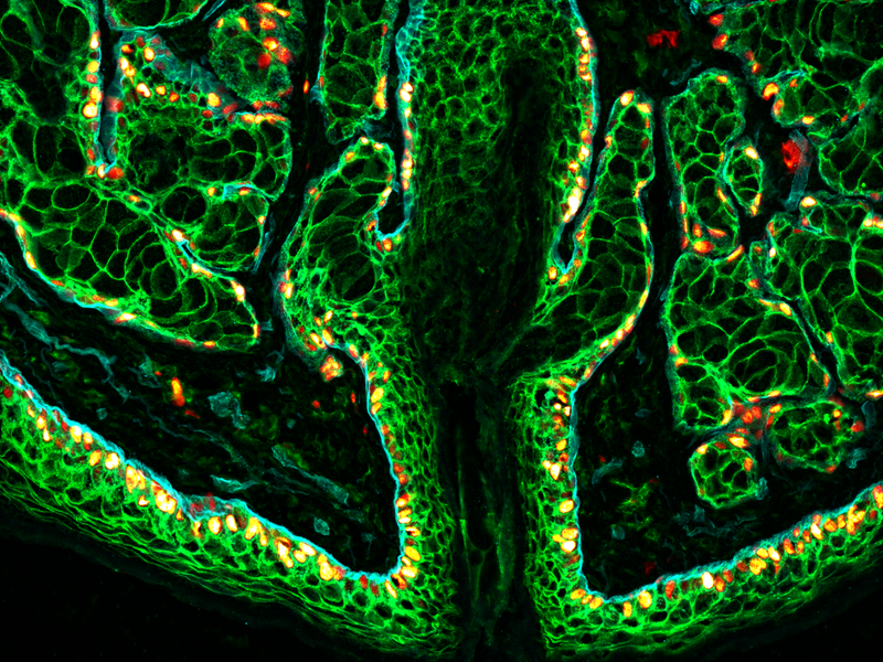
Title: My little green cactus (Mein kleiner grüner Kacktus)
The immunofluorescence staining image captures a murine hair follicle accompanied by sebaceous glands, with the stained proteins E-cadherin (green), Ki67 (red to white), and ITGa6 (cyan) highlighted. This multi-color depiction offers a dynamic view of cellular interactions and activity within these structures. E-cadherin emphasizes cell adhesion, Ki67 marks proliferating cells, and ITGa6 reveals basal epithelial cells. The resulting image provides a comprehensive insight into the spatial distribution of these proteins. The image was taken using a benchtop Evos M5000 Imaging System (20x magnification, Thermo Fisher Scientific).
Nicole Michelle Schottmann
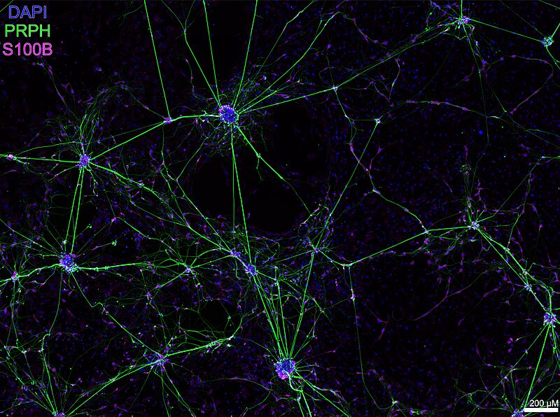
Title: Schrödinger’s Myelination
A culture of human induced pluripotent stem cell derived sensory neurons (iSN) after magnetic activated cell sorting (MACS).
MACS was used to reduce the amount of cells of unknown origin (feeder cells) differentiating alongside the iSN culture for a more specific in vitro model. After MACS, the feeder cells show a more homogenous morphology with some cells seemingly in a myelination-like contact with neurites of the iSN. TileScan acquired using the inverse microscope DMi8, Leica, Wetzlar, Germany.
Abbreviations: DAPI: 4′,6-Diamidin-2-phenylindol, : PRPH: Peripherin, S100B: S-100 Protein Subunit Beta
Trushnal Waghmare
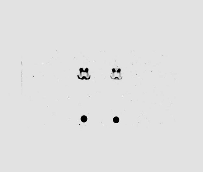
Title: Ceramide moustache ghost
Neutral sphingomylinease activity was measured in CHO cells.
Thin layered chromatography was performed to determine the enzyme activity from sphingomyelin (dot, bottom) conversation to ceramide. The converted ceramide looks like scary two eyes with a moustache. The image was captured on the LICOR Odyssey imaging system and analysed using Image Studio software.
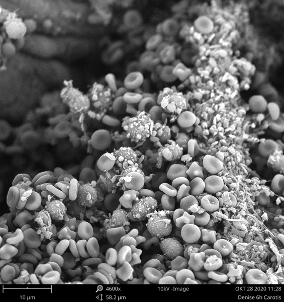
Denise Johnson
Title: Inside a thrombus
Acute venous and arterial thromboses are the most common cause of death in developed countries. The most important diseases to be mentioned here are myocardial infarct and stroke, for which the mortality is unacceptably high despite advances in research.
But how does a thrombus look like? By scanning electron microscopy, we investigated the components of thrombi induced by mechanical injury of the mouse Arteria carotis communis. On the image, red blood cells, platelets and leukocytes are trapped in a fibrin mesh (strands), forming the blood clot.
Tuğçe Çimen
Title: Cross talk between neutrophils and platelets in the inflamed microcirculation.
This confocal microscopy image shows blood vessel walls (endothelial cells (CD31 staining, blue) and pericytes (alpha-SMA staining, red) in an inflamed mouse cremaster muscle (LPS treatment). Luminal neutrophils (MRP14 staining, green) that have adhered to the vessel wall extensively interact with platelets (GPIX staining, white). With my research, I aim to determine the mechanisms by which platelets help circulating neutrophils to breach blood vessel walls and enter extravascular sites during inflammatory responses in vivo.

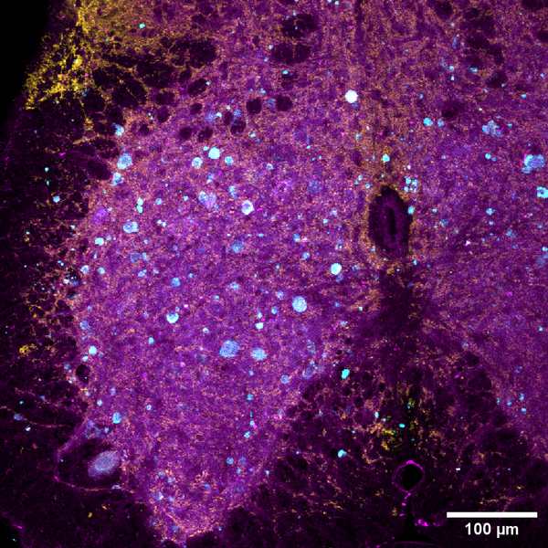
Alexander Veh
Title: Help me I’m swelling!
Confocal image of sacral spinal cord from Plekhg5 deficient mice. Plekhg5 causes deficiency in autophagy resulting in accumulation of synaptic vesicles (SVs) positive for Atg9 in axons of motoneurons. Accumulation of SVs leads to motoneuron death resulting in neurodegenerative diseases. SVs are positive for transmembrane marker Atg9, lysosomal marker Lamp1 and SV marker Synaptophysin. Immunohistochemistry staining with antibodies against Atg9 in cyan, Synaptophysin in yellow and Lamp1 in magenta. Z-stacked image with 20x magnification scanned with an Olympus FV1000 confocal microscope; scale bar 100 µm.
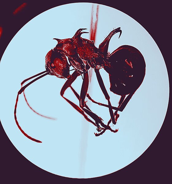
Vibhuti Bhat
Title: My EUREKA! moment
This is a Formicinae ant species called Polyrachis bellicosa. It is found in the South East Asian island countries of Philippines, Malaysia and Papua New Guinea (PNG).
I collected it with forceps and a falcon tube during field sampling in PNG. It's a literal Eureka moment for me because it's a hard find!
The unique hook shaped spine on the propodeum and the beautiful red coloured mesosoma differentiates it from the other ant species in the Polyrachis Genus.
I mounted the ant on a pinning block and took this picture with my phone camera while observing it under the microscope.
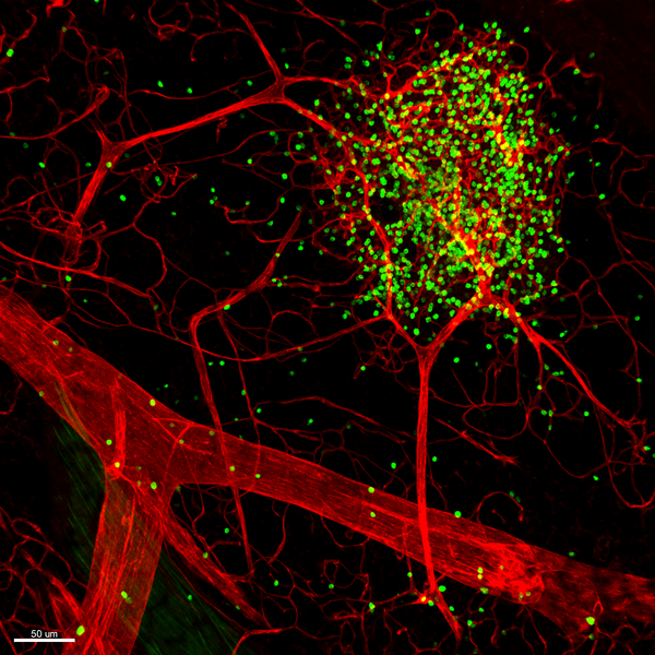
Ye Ouyang
Title: Tree
This pictures was captured from mouse mesentery with Leica SP8 confocal microscope. Vascular endothelial cells are shown in red and lymphocytes are shown in green. The lymphocytes formed tertiary lymphoid organ like cluster, and may play an important role in immune defence.
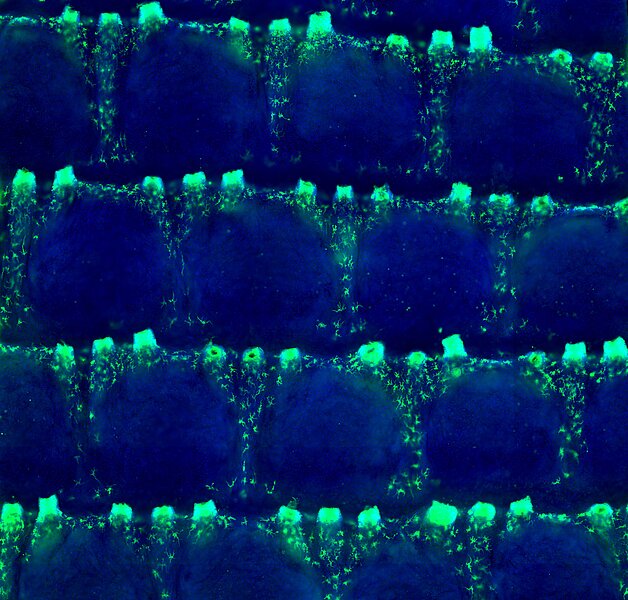
Muthita Khamwong
Title: Honeycomb architecture of Langerhans cell network in murine tail
The picture shows Langerhans cells (LC), immune sentinels privileged located to scan environmental antigens to trigger immune responses, on the mouse tail epidermis.
In contrast to homogenous network of LC in the murine ear and back epidermis, the LC locate exclusively in the specific domain on mouse tail called “interscale” and nearby triplet unit of hair follicles, creating an immune-privileged area called “scale” zone.
LC network forms ring structures surrounding the immune privileged zone to craft the honeycomb-like architecture on the mouse tail epidermis.
The image was created by performing immunofluorescence staining of epidermal sheet. LC were stained with the specific marker EPCAM and visualized by OLYMPUS VS200 slide scanner.
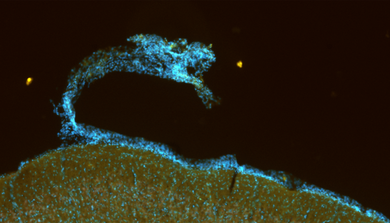
Martina Wilhelm
Title: Dragon of the Mind: Serendipitous Discovery in a Rat Brain Slice
Here a sagittal slice of a rat brain was stained after a viral injection into the cuneiform nucleus to trace ascending and descending glutamatergic structures. Tile imaging in Fluorescence microscopy makes it possible to display one large image, by “stitching together” various smaller images. This image consisting of 4 tiles is just a tiny section of the whole image consisting of 322 tiles. While watching the image grow tile after tile, we were greeted by this tiny dragon on the frontal cortex of the brain slice.
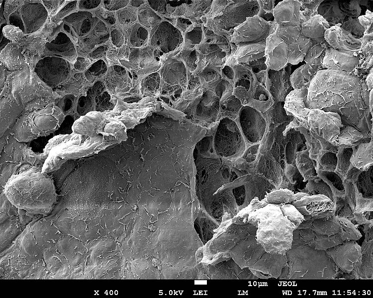
Sebastian Haeusner
Title: Into the Honeycomb!
Articular cartilage defects are widespread diseases with currently limited therapeutic options to restore cartilage function and integrity. Defects in cartilage architecture can lead to articular joint inflammation, pain and cartilage degradation, well known symptoms of osteoarthritis. In an approach to restore articular cartilage within the EU-funded BIO-CHIP project, autologous human nasal chondrocytes were isolated and in vitro cultured on a collagen matrix for reimplantation purposes. In order to assess maturation state, microarchitecture and ultrastructure during a good laboratory practice (GLP) compliant study, the in vitro engineered cartilage was imaged by scanning electron microscopy. The native ultrastructure was preserved using freeze cracking with liquid nitrogen to reveal the inner layers of the specimen. The image at hand shows the very top layer of cartilage, the so called superficial zone that consists of flat ellipsoid chondrocytes. During freeze cracking, some layers peeled off, revealing the underlying cavities that compose a „honeycomb like structure“ of chondrocyte capsules (lacunae). On the lower left side, a single chondrocyte can be observed that was dragged from its lacunae during sample preparation. The unique microarchitecture of articular cartilage is responsible for keeping the joints healthy every step of the day, carrying the weight of the human body during a whole lifetime.
Image taken on a JEOL Scanning Electron Microscope, at 400X, 5kv, scale bar 10μm
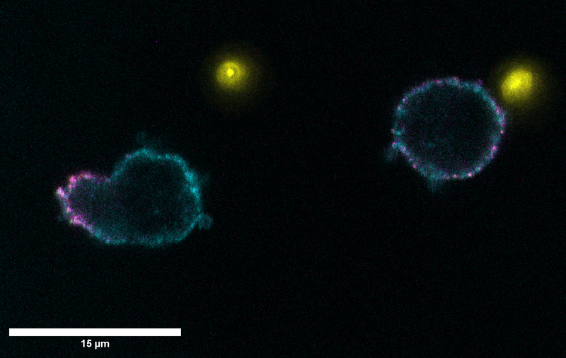
Bastian Just
Title: The dark side of the moon (or the neutrophil)
While blunted platelet reactivity and decreased platelet count would indicate otherwise, one major complication of sepsis is the increased risk for thromboembolic events. The induction of thrombotic events during inflammation is termed immunothrombosis and while neutrophils might be of high importance for bacterial clearance during infection, they themselves can trigger thrombotic events by neutrophil extracellular traps. By confocal laser scanning microscopy (Leica SP8) it can be observed that during sepsis neutrophils (in cyan by anti-CD15-APC) exhibit a clustering of P-selectin glycoprotein ligand-1 (PSGL-1) (in magenta by anti-PSGL-1-SB600), which facilitates the increased interaction with platelets (yellow by anti-CD42a-BV421) and reciprocal recruitment of the neutrophils, leading to enhanced thrombus growth in sepsis. Blocking of leukocyte PSGL-1 could allow for anti-thrombotic treatment without negatively influencing haemostasis.